
HSC Biology Syllabus Notes
Module 5 / Inquiry Question 2
Overview of Week 2’s Inquiry Question
Learning Objective #1 – Model the processes involved in cell replication, namely mitosis
Learning Objective #2 – Model the processes involved in cell replication, namely meiosis
Learning Objective #3 – Model DNA replication using the Watson and Crick DNA model, including nucleotide composition, pairing and bonding
Learning Objective #4 – Assess the effects of the cell replication processes on the continuity of species.
EXTRA Learning Objective #5 – History behind the Watson and Crick DNA model (Coming Soon)
NEW HSC Biology Syllabus Video – Cell Replication
Week 2 Homework Questions
Week 2 Curveball Questions
Week 2 Extension Questions
Solutions to Week 2 Questions
Overview of Week 2 Inquiry Question
WELCOME BACK to Week 2 of your Year 12 HSC Biology Syllabus Notes!
Well done for sticking around! You will do well, keep it up!
In this week’s notes, we will explore the mechanisms of mitosis, meiosis and the process of DNA replication that occurs during the two types of cell division.
In last week’s notes, we touched on Darwin’s Theory of Evolution by Natural Selection and how reproduction plays an important role in it. We also learned about how Darwin’s Theory and, hence reproduction, is important to the continuity of species!
However, up until now, we have only briefly mentioned the terms mitosis and meiosis under the categories of asexual and sexual reproduction respectively. So, in this week’s notes, we will dive deeper and explore how mitosis and meiosis actually works!
Remember in last week’s notes, we learned that part of Darwin’s Theory of Evolution states that there is a variation in the genetic material (gene pool) of a population? Well, there are different mechanisms in meiosis that give rise to such genetic variation in the population! Let’s move on to see how!
Oh yes, one last thing, the overarching inquiry question for this week’s learning objective is the following:
How important is it for genetic material to be replicated exactly?
We will using what we learned from the learning objectives to answer this inquiry question at the end!
Learning Objective #1 - Model the processes involved in cell replication, namely mitosis
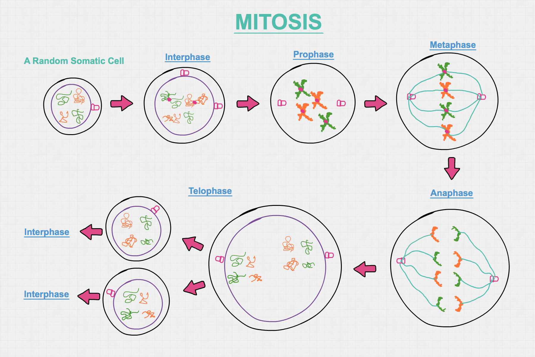
The following diagram illustrates the processes involved in mitosis
There are two terms that you need to know before we get start understanding the diagram! These terms are:
Chromatid: A single-stranded chromosome
Chromosome: A molecule that is made up of DNA and protein
Some terms you already know from Preliminary HSC Biology:
Nuclear membrane (depicted as the purple circle in diagram)
Cell membrane (depicted as the black circle in diagram)
Now that we know learnt some new terminologies, let’s explore what is exactly occurring in each of the stages of the mitosis illustrated in the diagram above!
Random Somatic Cell: Mitosis starts off with a somatic (body) cell, i.e. a cell that is not involved in the production of gametes (Gametes can be sperm or egg cells). The somatic cell is a diploid cell. A diploid cell means that it has two sets of each chromosome. For example, in humans, we have 23 sets of chromosomes. Each set of chromosome contains 2 chromosomes that are homologous to each other. We call them homologous pairs. We will explore what homologous pairs actually mean later as we get into the steps of mitosis. A haploid cell only contains one set of each chromosome, i.e. contain only half the amount of total chromosomes compared to diploid cells. There is only one chromosome per ‘set’.
At the end mitosis, the number of chromosomes is retained. This means that, at the conclusion of mitosis, each of the two daughter cells that are produced from the parent somatic cell are also diploid cells.
Interphase (Step 1): DNA replication occurs here. Each chromatid (single stranded chromosome) has its DNA duplicated, forming another chromatid that is genetically identical. These two genetically identical chromatids are called sister chromatids. Remember a chromatid is a single-stranded chromosome and as stated earlier in the definitions. Also note that a chromosome is made up of DNA and protein.
I have coloured the chromatids in the above diagram to outline that there are two sets of chromosomes (orange and green sets), i.e. two homologous pairs. In reality, humans have 23 sets or homologous pairs but only two pairs are depicted for the purpose of simple illustration.
It is important to note after DNA replication, the number of chromosomes have not changed!
There are still two sets of pairs illustrated in the somatic cell after interphase! The chromosomes have just changed from being single stranded (chromatids) to double stranded (sister chromatids).
So, after DNA replication, there are still 23 sets of chromosomes in humans like there is 2 sets of chromosomes illustrated in the diagram.
The following illustration depicts how the number of chromosomes have not changed after DNA replication.

Apart from the DNA being duplicated, the centrosomes (illustrated as the two pink ‘rectangles’ at right angles) have also been duplicated during interphase. Each of the rectangles represents a centriole in the centrosome if you are curious. Centrosomes play an important role in the later stages of mitosis.
Prophase: During prophase, the chromosomes coil up. You can now see chromosomes in their classic “X” shape under a compound or light microscope. During prophase, the nuclear membrane dissolves in the cytoplasm. Also, the centrosomes begins to move and align up at opposite ends of the cell’s equator
Metaphase: During metaphase, the chromosomes line up above each other along the poles the cell. The microtubules (fibres structures illustrated as blue lines), which attached to the centrosomes, will now have access and attach to the chromosomes’ centromeres (the point of which the sister chromatids in each chromosomes are attached, illustrated by the pink dot). The microtubules randomly attaches to the chromosomes’ centromeres. So, microtubules effectively join centrosomes and chromosomes together.
Anaphase: During Anaphase, the chromatids that are attached to centrosomes via microtubules are being pulled towards opposite sides of the somatic cell. The cell membrane is also starting to alter its shape for cell division.
Telophase: During Telophase, single-stranded coiled chromosomes start to uncoil. Cytokinesis occurs and the nuclear membrane starts to form again. The somatic cell divides into two. Each daughter cell have identical and equal amounts of genetic material as the original parent somatic cell. Each daughter cell is capable of entering interphase to undergo mitosis when given instruction to do so. No genetic variation created.
Having explored the mechanisms of the asexual reproduction process, Mitosis, we will now move on to explore meiosis, a sexual reproduction process.
There are two stages of Meiosis because there are two sets of cell division. In mitosis, one cell split into two. In meiosis, one cell splits into two and each of the two cells further splits into two. So, at the end of meiosis, there are four cells (gametes) produced.
Learning Objective #2 - Model the processes involved in cell replication - Meiosis I
Sexual Reproduction - Meiosis I

Unlike Mitosis, Meiosis starts off with a germ cell rather than a somatic cell. A germ cell is found in the reproductive organ of an organism can undergo meiosis to produce gametes such as sperm and egg cells, depending on the gender of the organism.
Like a somatic cell, a germ cell is also a diploid cell. However, unlike mitosis, meiosis does not maintain the overall number of chromosome number throughout the process. Therefore, the gametes cells that are produced at the end of meiosis are NOT diploid but haploid cells.
Interphase I: DNA replication occurs here. Each chromatid has its DNA duplicated, forming another genetically identical (sister) chromatid. I have coloured the chromatids here. The yellow chromatids are from the father and the green chromatids are from the mother.
Similar to Mitosis’s interphase stage, the number of chromosomes have not changed before and after interphase. Apart from the chromatids, the centrosomes have also been duplicated.
Prophase I: During prophase, the chromosomes coil up. The nuclear membrane dissolves in the cytoplasm. The centrosomes begin to move and align up at opposite ends of the cell’s equator. You can now see chromosomes under the microscope in their classic “X” shape. Unlike in mitosis, homologous chromosomes in propose of meiosis will line up side-by-side (not on top of each other) across the equator of the cell for crossing over.
During crossing over in Prophase I, the double-stranded homologous chromosome pairs (one from father and one from mother) exchange their genetic material. This will mean that the (non-sister) chromatids involved in the crossing over would create new allele combinations. This means that the resulting gametes can inherit new allele combinations that are different from their parents.
Illustration of Crossing Over

Illustration of new allele combinations being created

Notice that PRIOR to crossing over:
The allele combinations in each of the two double chromatids were (BHC and BHC).
The allele combinations in each of the two pink chromatids were (bhc and bhc).
Notice that AFTER crossing over:
As seen in the above diagram, the allele combinations for the chromatids (from left to right): BHC, bHC, Bhc and bhc.
Two new allele combinations are created. These are bHC and Bhc which did NOT exist before crossing over or if crossing over did not happen.
Metaphase I: As the nuclear membrane dissolves, the microtubules attached to the centrosome can bind with the chromosome at their centromeres. This binding process of microtubules to chromosomes is random. This random binding process results in what is called the independent assortment of non-homologous chromosomes.
Independent assortment is the process where the alleles specifying for different genes (in non-homologous chromosomes) assort themselves independently. This would therefore mean the independent alignment of the chromosomes between non-homologous pairs on the equator of the cell. This process of independent assortment will affect the genetic material of the two haploid cells that will be produced in the later steps.
We will explain how independent assortment increases genetic variation very shortly after explaining Meiosis I and Meiosis II.
Illustration of Independent Assortment

Anaphase I: The microtubules move the chromosomes in each homologous pair move to different sides of the cell membrane. As the microtubules do no selectively bind to a chromosome (as mentioned in Metaphase I), the side of the cell to which the chromosomes will be pulled towards will depend on how they are connected to a centrosome via microtubules.
Telophase I: The coiled chromatids of each chromosome starts to uncoil. The microtubules begin to break down and a new nuclear membrane is created to enclose the chromosomes. Since each chromosome of the homologous pair are now in different cells, there is no longer homologous pairs in each of the haploid cell. This is all due to independent assortment in Metaphase I.
Sexual Reproduction - Meiosis II
Meiosis I has been successful! Let’s complete the germ cell’s division in Meiosis II!

Prophase II: Centrosome duplicates for each haploid cell. Chromosomes coils up. The nuclear membrane formed during the Telophase I dissolves.
Metaphase II: The two centrosomes in each of the haploid cells move opposite poles of the cell. The chromosomes line up side-by-side along the equator of the cell. Microtubules attaches the chromosomes to centrosomes.
Anaphase II: Random segregation occurs here. The process of random segregation refers to the random separation of chromatids to different poles in a haploid cell and, ultimately, affects the chromatids that end up in each of four gametes.
Note that each chromatid may contain different alleles for a particular gene which end up in different gametes.
During Anaphase II, the microtubules separate the sister chromatids of each chromosome, pulling one chromatid to a different pole in each haploid cell. The result is that on each pole for each haploid cell, there are two chromatids. This segregation process is random, i.e. you cannot determine which chromatid will end up at which of the four gametes during Cytokinesis II which occurs in the next step (Telophase II). Unlike independent assortment, we are dealing with individual chromatids here instead of homologous chromosome pairs. Segregation increases the genetic variation of the gametes and thus offspring (derived from gametes). We will clarify as to why segregation increases genetic variation shortly.
Telophase II: The coiled chromatids starts to uncoiled. Cytokinesis II occurs during Telophase II, forming four haploid gamete from the splitting of each of the two haploid cells. Each of the gamete inherits one allele of every gene. This is because each of the double-stranded chromosomes that contains two alleles for different genes have separated during Anaphase II.
During Telophase II, the microtubules begin to break down and a new nuclear membrane is created to enclose the two chromatids in each of the four daughter cells (gametes). One centrosome to each daughter cell. Depending on the germ cell and, hence gender of the organism, the four gamete is either sperm or egg cells. The gamete can fuse with its opposite kind (e.g. sperm cell with egg cell or vice versa) to form a diploid cell, a zygote so that the zygote will have two alleles for a given gene.
Difference between Independent Assortment and Random Segregation
Independent Assortment and Random Segregation occur in different stages of Meiosis
Crossing over, independent assortment and random segregation does not apply to Mitosis.
Independent Assortment occurs during Metaphase I of Meiosis I
Independent assortment is the process where the alleles specifying for different genes (in non-homologous chromosomes) assort themselves independently.
Random Segregation of chromatids (chromosomes) occurs in Anaphase II of Meiosis II
Independent Assortment in Metaphase I results in the separation of double-stranded CHROMOSOMES from their homologous pairs in Anaphase I.
Random Segregation deals with separating CHROMATIDS of EACH double-stranded chromosome.
So, effectively, independent assortment deals with sorting of alleles of different genes in non-homologous chromosomes and random segregation deals with splitting of double-stranded chromosomes to single-stranded chromatids (where each chromatid may have different alleles for a particular gene).
How does Independent Assortment and Random Segregation increase genetic variation?
If you recall, the sister chromatids in double-stranded chromosomes in meiosis II have different alleles (due to crossing over during Prophase I in Meiosis I). You can look at Anaphase II in the Meiosis II diagram where one chromatid both has both green and orange alkene due to crossing over. Thus, the separation of sister chromatids from each double-stranded chromosome in Anaphase II will increase genetic variation by segregating one chromatid that may or may not have undergone crossing over to different poles of each haploid cell which further divides into gametes during Cytokinesis II in Telophase II.
Therefore, the resulting gamete may inherit different alleles for different genes.
Keep in mind that during the segregation process in Anaphase II prepares for each gamete to inherit one allele for each gene from the parent, which is why it is a haploid. Upon fertilisation, two gametes (each with one allele for a particular gene) will combine so that the formed zygote will then have two alleles for a given gene, i.e. diploid.
For independent assortment, it is the process where alleles specifying for different genes in non-homologous pairs will assort themselves independently during metaphase I. This is because the double-stranded chromosomes in their non-homologous pairs can be aligned independently to the other non-homologous pair which will increase genetic variation for the two haploid cells formed in cytokinesis I. Therefore, independent assortment will determine the alleles for different genes such each of the two haploid cells can inherit which, ultimately, affects the allele for each gene that each gamete can inherit at the end of Cytokinesis II in Telophase II.
Learning Objective #3 - Model the process of DNA replication using Watson and Crick DNA Model, including the nucleotide composition, pairing and bonding

Recall that the DNA of chromatids were duplicated during Interphase of Mitosis and Interphase I of Meiosis I.
How did the DNA of chromatids exactly duplicate? Well, this is known as DNA replication. We will now explore the Watson and Crick DNA model and see how did the DNA duplicate during mitosis and meiosis.
DNA replication occurs in the nuclear membrane. If you return to mitosis and meiosis diagrams, you can see that the nuclear membrane is depicted as a purple circle.
Before we can start to learn how DNA replication actually works, we should first learn some new definitions.
A polymer is a substance that is made up of many repeating units called monomers. DNA is a polymer and it is made up of many repeating monomer units called nucleotides.
A nucleotide (monomer) has three main components. Each nucleotide has a deoxyribose sugar molecule, a phosphate molecule (negatively charged) and one of the four nitrogenous base (adenine (A), thymine (T), guanine (G) or cytosine (C)).
As you see in the diagram below, the nitrogenous bases are joined together by chemical bonds. Adenine joins with Thymine through two chemical bonds. Guanine and cytosine join together through three chemical bonds. Nitrogenous bases join together via complementary base pairing. This means that, under normal conditions, Adenine only bonds with Thymine and Cytosine only bonds with Guanine.
We did just mention that a nucleotide contains a deoxyribose sugar molecule and a phosphate molecule. In the diagram below, the ‘backbone’ of the two DNA strands, depicted as blue lines, is composed of ALTERNATING phosphate and deoxyribose sugar molecules. Phosphate, Sugar, Phosphate, Sugar, etc. Of course, they are joined together via chemical bonds too.
The diagram above is a detailed diagram of a DNA helix that has been unwound and showing the sugar-phosphate backbone on both strands.
Notice the nucleotide molecule under ‘Legend’. Depending on the nucleotide, the nitrogenous base can either be adenine, thymine, cytosine or guanine. I just happened to depict adenine.
The full name for DNA is deoxyribonucleic acid.
The process of DNA Replication

Step 1: DNA replication starts with a double-stranded DNA helix molecule.
Step 2: The enzyme, helicase, attaches to and unwinds the double-stranded DNA helix. Helicase also facilitates the breaking of hydrogen bonds between the nitrogenous bases (Adenine, Thymine, Guanine and Cytosine). This would lead to the separation of the two DNA strands.
Step 3: Each of the two now separated DNA single strands act as templates for free (available) nucleotides from the nucleoplasm (inside nuclear membrane) to join via complementary base pairing, i.e. Adenine bonds with Thymine and Guanine bonds with Cytosine. The enzyme, DNA polymerase, moves along the DNA strands during this process to catalyse the reaction, allowing complementary base pairing to occur.
Step 4: The enzyme, DNA ligase, secures each of the new DNA strands formed with free nucleotides (monomers) with complementary base pairing.
Step 5: Each DNA double strands return to their chemical stable state by winding up spontaneously to form a two double-stranded DNA helix.
Learning Objective #4 - Assess the effect of cell replication processes on the continuity of species
If an organism has favourable characteristics that allow it to tolerate the selective pressures of its ambient environment, cell replication is critical in allowing such favourable characteristics to be passed onto offsprings. With the offspring inheriting favourable adaptations, they would have a higher chance of survival in its environment than without. This is why mitosis is important. Mitosis basically produces clones, thus ensuring that favourable characteristics are inherited by offsprings throughout the population. This is seen in plants’ runners leading to new plants that are clones (genetically identical) of their parents.
In humans, our muscle cells, hair cells and so many other cells reproduce via mitosis. Mitosis is important for cell development and growth!

Compared to meiosis, there is no crossing over in mitosis. Crossing over occurs during Prophase I of meiosis where non-sister chromatids in homologous pairs exchange genetic materials, creating new allele combinations.
As crossing over increases the genetic variation in the resulting offspring and, thus the species population, it reduces the probability that a sudden change in environment will lead to an extinction event. This will therefore ‘ensure’ the continuity of a species through meiosis.
In addition to crossing over in meiosis, the processes of independent assortment and random segregation during meiosis also help increase variation in the resulting offspring. In general, the both lead to increased genetic variation of the resulting gametes. We have already touched on this during the meiosis steps but let’s review and tie it to the learning objective!
Similar to crossing over, we have already talked about independent assortment and random segregation already in terms of increasing genetic variation in the gametes and, thus, offspring when the gamete is fertilised. Therefore, all of these processes (including random fertilisation) serves to increase the genetic variation in offspring. By increasing genetic variation, the offspring of the next generation would have different characteristics which would increase the probability of the species’ population surviving the event of a sudden change in the ambient environment. This is because the survival of the species population of the next generation will not be dependent on a fixed characteristic or characteristics as variation increases.
Also, the unique allele combination in each of the four gametes (per germ cell’s meiosis) increases the genetic variation of the zygote that is formed. This is because any gamete have equal chance (25%) in combining with its counterpart gamete (e.g. sperm with egg or vice versa) to form a zygote. This would therefore increase the genetic variation in the offspring and thus ‘ensures’ the continuity of a species.
Summary - Broad answer to Week 2 Inquiry Question
The advantages when genetic material (DNA) is replicated exactly
Exact replication has the advantage in that it allows its parent(s)’s favourable characteristics to be passed on to offsprings. This is because it will allow the offsprings to have the favourable adaptations that enables it to tolerate its environment’s selective pressures.
The disadvantages when genetic material (DNA) is replicated exactly
It will not be ideal for a parent with unfavourable characteristics to be passing on undesirable characteristics (allele combinations) to offsprings via mitosis. Therefore replicating DNA exactly will be bad in this situation.
Also, even if all of the parents’ characteristics that are passed down to offsprings are favourable, exact replication of genetic would decrease genetic variation in a population. So, in the event of a sudden change for a species’ ambient environment, there may be a threat of extinction.
Week 2 Homework Questions
Week 2 Homework Question #1 – Crossing over occurs in both mitosis and meiosis. True or False? Explain your answer. HINT: ‘A clone has no genetic variation from their parent’ (2 marks)
Week 2 Homework Question #2 – Describe the process of crossing over in terms of where, when and how does it occur (4 marks)
Week 2 Homework Question #3 – Describe the process of independent assortment in terms of where, when and how does it occur (4 marks)
Week 2 Homework Question #4 – Describe the process of random segregation in terms of where, when and how does it occur (4 marks)
Week 2 Homework Question #5 – Explain how does crossing over, independent assortment and random segregation affect the genetic variation of an offspring (6 marks)
Week 2 Homework Question #6 – Describe the process of DNA replication, include diagrams in your answer (6 marks)
Week 2 Homework Question #7 – Explain the relationship between DNA replication, mitosis and meiosis (4 marks)
Week 2 Homework Question #8 – How does DNA replication affect genetic variation? (4 marks)
Week 2 Homework Question #9 – Suppose a DNA strand is made up of 16% Thymine. What is the percentage composition of Adenine, Guanine and Cytosine in the same DNA strand? (3 marks)
Week 2 Homework Question #10 – How does Meiosis and Mitosis help support Darwin’s Theory of Evolution? (8 marks)
Notice how you can now provide a more comprehensive answer to Question 2 in Week 1’s Homework set.
Week 2 Curveball Questions
Curveball Question 1 – How does meiosis and mitosis help ‘ensure’ the continuity of a species? (6 marks)
Curveball Question 2 – What are the advantage and disadvantages of genetic material being replicated exactly? (4 marks)
Curveball Question 3 – How does random segregation, crossing and independent assortment ‘ensure’ the continuity of a species? (6 marks)
Curveball Question 4 – Explain the difference between natural selection and survival of the fittest? (4 marks)
Curveball Question 5 – Explain the importance of cell development and growth (6 marks)